

1 Introduction
Brain images, whether digital, electronic or illustrated, are pervasive in North American life. Neuroscientists, neurologists, neuropsychologists, and other health professionals use them when communicating about brain pathologies, emotional reactions, and personality traits in research and clinical settings [Dumit, 2004]. They are employed to convey scientific information in conference papers, scientific publications, public health infographics, and bioscientific websites [Joyce, 2006]. The more ‘scientific’ an image appears, the more authority it has to communicate facts about brain health, human behavior, personhood, and cognition, since biomedical images denote objectivity and expertise [Beaulieu, 2001; Gruber, 2021; McCabe & Castel, 2008].
Brain images also circulate beyond the professional health realm, appearing in courtrooms when investigating self-determination in criminal cases and in the media when guiding study plans, social media habits, dietary choices, and other daily practices [Dumit, 2004, 2014]. Likewise, they are featured in films, art pieces, and cartoons for their entertainment value in popular culture and are used as appealing elements in scientific news for their aesthetic qualities [Khalili-Mahani & Loos, 2023]. In this way, brain images are technical tools that attract and influence how we understand ourselves, our bodies, and our interactions with others, operating across expert and nonexpert domains [Sturken & Cartwright, 2018].
In this article, we examine the brain images used to communicate neuroscientific knowledge through digital media, recognizing that these images are both social and technical. This study is part of a larger project exploring the social representations of brains. Specifically, we are interested in how brain images influence realities, determining which realities are deemed important and which are not. For this purpose, we analyze the brain images featured on a website that promotes the use of a neuropsychological test, the Montreal Cognitive Assessment (MoCA). The MoCA test is considered one of the leading standards to screen mild cognitive impairments related to medical conditions such as Alzheimer’s disease, Parkinson’s disease, and brain injuries. Clinicians and researchers administer it via various subscales to assess cognitive domains such as attention, concentration, and executive function. This is performed by completing tasks such as matching numbers with letters, identifying animals, drawing a clock, and recalling lists of words [Geller & Slicer, 2024].
As is the case with brain images, the MoCA test circulates between medical and public arenas, making it an object of political and cultural interest. For example, one of the preoccupations during the last United States Trump-Biden campaign, before Kamala Harris was the Democratic Party’s choice, was their age, since both candidates would be over 80 years old when in office. Opposing politicians used this as an opportunity to challenge the candidates’ capacities, prompting them to undertake assessments such as the MoCA to demonstrate the cognitive prowess required for such a powerful leadership position. These concerns were especially encouraged by Donald Trump and his team, who affirmed that he had taken the MoCA test in 2018 with ‘excellent’ (passing) results. In the meantime, Joe Biden refused to take this test, claiming that governing the US was the best demonstration of his cognitive functions. These political tensions sparked a public controversy, with health institutions and prominent academic figures, raising questions about the test’s potential and limitations. Within this controversy, Ziad Nasreddine (the MoCA’s original developer) and other experts suggested that Biden’s hesitations in public speeches made him a candidate for the MoCA [Ortega & Nirappil, 2024]. In these socio-political contexts, such medical protocols take on new meaning as they shape national and international conversations not only about brain health, but also about the ‘fitness’ of political leaders.
The MoCA’s official website is called MoCA Cognition. It is an open site that describes the history, uses, and innovations of the MoCA test. For different fees, it offers training for those interested in administering the MoCA test to patients, access to the latest MoCA applications, and software for interpreting test results. The target audience for this website includes clinicians, researchers, patients, members of medical associations, and policymakers. Therefore, this site has two main goals: first, to inform different audiences about cognitive testing; second, as a private initiative, to promote the use of the MoCA for profit. In this way, MoCA Cognition combines medical development and marketing, blurring the lines between scientific research, advertising, and business in the medical field.
Several websites are dedicated to promoting various cognitive tests, including the Cognitive Log (Cog-Log), the Oxford Cognitive Screening (OCS), the Rowland Universal Dementia Assessment Scale (RUDAS), and the Mini-Mental State Examination (MMSE). These websites typically incorporate tables with numbers, references to studies, recordings of conferences, videos of patients taking tests, and extensive written information. The MoCA Cognition website shares some of these features by displaying charts with statistics and professionals applying the MoCA to patients. However, unlike these other websites, MoCA Cognition uses a range of images to express its message, including for example, photographs of urban cities and high-technology facilities. Moreover, unlike other websites, it heavily relies on brain images as one of its primary communicative mechanisms. They appear as Magnetic Resonance Imaging scans (MRI), drawings, and digital displays. Yet, the MoCA test is not a neuroimaging tool, but a cognitive test; its outputs are not brain representations, but numbers plotted on range charts. Therefore, the images on this website serve a particular purpose related to the charisma of brain images, beyond merely representing cognition or standardized medical protocols.
The gap between the MoCA test as a cognitive tool and the brain images featured on the MoCA Cognition website highlights the relevance of examining how brain images communicate science in digital media. Consequently, we analyze these images to consider how they suggest ways of visualizing and understanding cognitive realities. To do this, we ask the following research question: what rhetorical, epistemological, and ontological work do brain images play on neuroscientific websites?
2 Theoretical frameworks
We begin this section by discussing the particularities of science communication through digital media and its connections to marketing and the depiction of body organs. Next, we explain the concept of the ‘rhetoric of science’ and show how it has been employed in the neurosciences. Lastly, we tackle the literature on brain visualization and the epistemological and ontological implications of current brain representations.
2.1 Digital science communication, marketing, and bodies
Recent approaches to science communication analyze a range of formats. They engage with both traditional media, such as scientific journals and books, and newer media, such as blogs, social media, scientific cafés, festivals, and scientific websites [Horst et al., 2017]. However, digital landscapes do not follow the same logic as traditional scientific media. Online science communication has fostered the public participation of multiple actors in the discussion of scientific matters, including politicians, non-governmental organization workers, and influencers. This opens avenues for increased interactions between scientists and the rest of civil society and, at the same time, entails risks related to the quality of the information being shared [Fähnrich et al., 2023]. Equally, private health websites facilitate the emergence of alternative ways of knowing that do not adhere to evidence-based medicine but are driven by profit. This has made the limits between consumption culture and medicine blurry [Erikainen et al., 2020].
One example of how marketing, digital science communication, and images are interconnected is the case of commercial milk formula. Rollins et al. [2023] argue that, despite the World Health Organization’s recommendations on breastfeeding and the proven adverse effects of commercial milk formula, marketing strategies have shaped the beliefs, political actions, and consumption habits of parents, communities, scientists, and policymakers. The industry’s marketing methods include messages that link this product to intelligence and early brain development. This includes AI-generated images of babies wearing glasses and playing with abacuses to symbolize their ability to read and count. However, there is no evidence supporting the connection between the development of these abilities and the consumption of commercial milk formula.
The way human organs are depicted in the media emphasizes certain realities while concealing others based on cultural preferences; for example, the frequent showing of ultrasound images in public when illustrating pregnancy [Sturken & Cartwright, 2018]. Ultrasound scans are widely regarded as ‘windows to wombs’, and the images they generate are seen as the first ‘pictures’ of babies, even though they are not direct visuals but data analysis outputs. Therefore, ultrasound images have helped foster a sense of fetal personhood by portraying fetuses as floating, separate entities, which blurs the pregnant bodies. As a result, they play a vital role in conversations about abortion and reproductive rights [Petchesky, 1987; Sturken & Cartwright, 2018].
A similar thesis has been sustained in Haraway’s [2013] concept of “god’s trick”, which posits that science presents its findings as if an omnipresent eye had obtained them, hiding any historical context or political implications. Therefore, even if anatomical images are presented as neutral pieces of evidence, they embody forms of political agency that shape our way of looking and acting in the world [Casini et al., 2022; Sturken & Cartwright, 2009, 2018]. Attuned to this discussion, we observe the images on the MoCA Cognition website, noting the flexibility of digital media in presenting scientific content, the connections between science and marketing, and the ways of looking encouraged by images of particular body parts.
2.2 Rhetoric of science
Traditionally, rhetoric has been linked with dishonesty and trickery; however, it appears in various fields like politics, marketing, and science. Broadly, rhetoric is any organized effort to persuade social actors of the truth of claims about reality [Pinch, 1993]. Based on Bloor’s [1993] symmetry principle, Science and Technology Studies scholar Trevor Pinch [1993] advocates for a comparative rhetoric that highlights the common persuasion techniques used across diverse areas such as science and marketing. Following this idea, Pinch observed that scientists and salespeople use similar tactics, like humor and comparisons, to gain support for their claims. Scientists use rhetoric in writings, speeches, demonstrations, and visual displays to persuade other scientists, funders, and the public about the truth of their statements, helping them secure resources and credibility. This approach has been particularly effective when persuasion happens subtly [Sismondo, 2009].
Neuroscientists are among the actors who use rhetorical strategies in scientific communication. For example, Mantilla [2018] examines the case of Argentinian neuroscientists involved in diffusion practices. In this case, neuroscientists employed three rhetorical strategies to justify the inclusion of neuroscientific discussions in the public sphere: they maintained that public engagement in science would foment critical thinking, they argued that scientific institutions are a model of democracy from which society needs to learn, and they affirmed that health policymaking must be carried out based on neuroscientific knowledge. Drawing on these insights, we examine the images on the studied website paying attention to the realities they promote, the subtlety of the elements they incorporate to persuade viewers of their validity, and the explicit discourse accompanying the visual persuasion strategies.
2.3 Brain images as epistemological and ontological devices
The popularization of technologies like electrocardiograms, X-rays, ultrasounds, MRIs, and an extensive array of visual medical devices has contributed to establishing the idea that the interior of the body is accessible, even without opening it. van Dijck [2005] calls this phenomenon “the transparent body”, a particular form of looking that assumes that the body is an entity susceptible to being unveiled by the eye. Further, the public discourse about neuroimaging has employed certain metaphors that obscure the epistemological problems implicated in the construction of medical imaging. In this vein, Carusi and Hoel [2015] argue against the photograph and window metaphors to describe neuroimaging as entries to the brain because they resemble a neutral position of the observer. On the contrary, these scholars sustain that science constructs objects that are not equivalent representations of nature.
Although neuroimaging appears relatively straightforward to the public, its construction is complex and uncertain. Dumit’s [2004] account of Positron Emission Tomography (PET) exemplifies one of the core paradoxes associated with neuroimaging. On the one hand, PET involves the collaborative work of several professions, difficulties in establishing clear parameters for normal and abnormal subjects, the preparation of radioactive molecules, and the work of algorithms to transform data into three-dimensional maps. On the other hand, the PET’s output appears to the public as elementary, transparent, clean, and colourful; almost like a snapshot. Similarly, Casini [2021] stresses the importance of aesthetics, affectivity, and craft practice in developing biomedical imagining. Then, diagnostic imaging is a blend of scientific and artistic knowledge that creates new worlds and alters the way we perceive existing ones. This can be noted in digital neuroimaging, where defined colors are implemented to maximize the visual contrasts building on minor data differences [Dumit, 2014].
Inspired by these conversations, we examine the images on the MoCA Cognition website, analyzing the epistemological problems they highlight or obscure; for example, how images depict various tools, such as MRIs and the MoCA test, as means to make the brain more accessible. Likewise, we are drawing on the insights of scholars affiliated with the ontological turn [Law, 2004; Martínez-Medina, 2022; Mol, 2002], who argue that the methods and representations scientists use shape realities and bring objects into being. In this sense, we examine the type of realities that are enacted by the studied images; for instance, whether they privilege simplicity or messiness when representing brains. Equally, we reflect on the ontological consequences of implementing aesthetic work when depicting brain structures.
3 Methods
Our method employs a variant of discourse analysis that focuses on images [Rose, 2023], which is refered to as Visual Discourse Analysis. This method examines how statements and images structure the way phenomena are understood and the subsequent actions that emerge from that understanding. A key tenet of this method recognizes that statements in the form of text and images have rhetorical effects that configure reality. Similarly, it identifies images as forms of discourse because they evidence and obscure objects according to cultural contexts.
One of the core ideas of Visual Discourse Analysis is intertextuality. According to Rose [2023], intertextuality recognizes that together verbal, textual, and visual media express discourses. On this basis, we examined the images displayed on the MoCA Cognition website, along with their accompanying signifying text descriptions. Equally, we engaged with the videos posted on it, paying attention to the image sequences and the spoken and written speech shown while they were played.
Overall, our analysis unfolded in an in-between place; it neither interpreted the developer’s intentions nor the appropriation and transformations experienced by the audiences during its use. Instead, we treated images and videos as informants in their own right, exploring their agency and acknowledging that they are not simply ornamental elements accompanying scientific communication [Rose, 2023]. What is more, sometimes, they lined up with textual messages, but in others, they contradicted and surpassed them.
3.1 Data collection
To collect our data, we browsed all sections of the MoCA Cognition website over 6 months in 2023. As a form of ethnographic participant observation, the first author also completed the online MoCA training, which offered a participant’s point of view, not to mention access to parts of the website that are not open to the public. This process involved capturing screenshots of all the images posted on the website, along with their accompanying text. After this, we downloaded the videos and the brochure. As a result, we collected 31 images and five videos. The videos included the main promotional video, the training video, and the videos dedicated to the new MoCA applications (MoCA Duo, MoCA SOLO, XPRESSO).
3.2 Data analysis
For the analysis, we followed the guidelines of Visual Discourse Analysis [Rose, 2023], concentrating on three themes: 1) the uses of brain images as rhetorical devices, 2) the ways in which the website’s images render brains and cognition as knowable entities, and 3) the realities these images put at the forefront.
The procedure went as follows: first, we observed all the images, texts, and videos several times, and recorded our initial impressions in analytical memos. Second, we prepared the materials for deeper analysis, labelling them by their placement on the website and with keywords related to their content. In addition, the video scenes were described in detail, and the verbal and written messages in them were transcribed. This information was placed in tables, as shown in Table 1, where we describe the last scenes of one of the videos. Third, we performed open coding, yielding 84 codes, most of which were accompanied by analytical notes. When coding, we first focused on the visual components and then considered their relationships with the textual and verbal content. The coding moved back and forth between our database and the website, which allowed us to examine each piece individually and in relation to the rest of the website’s interface. Fourth, we noted emerging themes, such as the presence of images combining human faces with cybernetic components. Fifth, we filtered out the codes and themes unrelated to the research questions by including only those clearly related to brain images and excluding those that addressed other issues. For instance, we excluded codes related to the medicalization of everyday life. We ended up with 21 codes and five themes, the latter corresponding to each section of the results (MRIs, brain drawings, binary brains, human-cybernetic brains, and colorful brains). Finally, we established connections among the remaining codes and themes.
|
Scene description |
Textual component |
|
The scene is set against a digital black background with white nodes and blue links, which quickly form a digital brain. Then it turns into a blue Earth globe that rapidly becomes a composed image: on the left, the actual world. On the right, a person is looking at it. |
Ziad Nasreddine in voice-over: MoCA cognition has the honor of being at the intersection of care, research, and digital innovation, with deep-rooted relationships around the globe. |
|
The take comes back to Nasreddine. |
Nasreddine: Let’s continue changing the landscape of cognition together. Popping message in white letters: TOGUETER |
4 Results
This section describes our findings according to five emerging themes: 1) the rhetoric involved in displaying MRI images; 2) the epistemological and ontological work done through brain drawings; 3) the gaze encouraged by movie-like digital images of the brain; 4) the hybridization between human faces and digital brains; and 5) the use of colors to produce differences and limits among brain structures. At this point, one clarification is needed. Our initial goal was to include images from the studied website along with our description to help the reader gain a sense of the work done by them; however, we were unable to secure copyright permissions to reproduce these images. As an alternative, we include images from open-access databases that are remarkably similar to the ones we analyzed, which hints at the current social imagination around brain images.
4.1 The MRI rhetoric
One of the most dominant rhetorical devices that emerges from our analyses is the use of brain MRI images. A brain on a laboratory bench would show a fatty, globular, rather ugly organ, but the MRI image transforms this into something at once beautiful and scientific. MRI images are often placed in the most visible parts of the MoCA Cognition website, occupying significant portions of the interface. This indicates their critical role in the overall messaging for their scientific authority and aesthetics [Casini, 2021].
One of the brain MRI images on the studied website is in the brochure. It is deployed on a tablet and introduces the heading “WHAT CAN MoCA DETECT?”. One hand is finger-pointing at the brain image on it. After this, there is a list of the cognitive domains the MoCA detects: “Short term memory”, “Visuospatial abilities”, “Executive functions”, “Attention, concentration, and working memory”, “Language”, and “Orientation to time and place” (see Figure 1).

MRIs assess the brain’s soft tissue, whereas the MoCA evaluates cognitive domains. Yet, the image suggests that the MoCA and MRI neuroimaging are closely connected. The MoCA exam and MRI are correlated but are not equivalent [Abou Elmaaty et al., 2019; Zhang et al., 2021]. Moreover, this correlation is not always straightforward since it implies certain degrees of probability, and, sometimes, the correlation is not positive. Further, there are marked difficulties in establishing direct links between brain imaging and neuropsychological variables by combining the results of devices that produce dissimilar objects such as these two [de Boer et al., 2020].
In this context, the MRI image featured in the brochure illustrates the flexibility of science communication through websites that serve both technical and marketing purposes [Erikainen et al., 2020]. There is a gap between the MoCA and the MRI. Still, it is bridged by pointing to a specific part of the MRI image to show that the cognitive domains detected by the MoCA occur in “visible” brain regions. In other words, a cognitive reality is presented, but it must be validated by its correlation with the MRI. Therefore, the image implies that MRI neuroimaging has a higher epistemic status than the MoCA, yet the brochure uses this status to reinforce the MoCA’s value.
Brain MRI images are also included in the training video. One of these images is projected onto a computer screen in a laboratory, as follows: a man wearing a white coat and a facemask looks at the screen while handling tubes containing a red substance and typing into the computer. An MRI brain image is being scanned in the middle of the screen while a message asks the video viewer, “WHY GET CERTIFIED?”
The appearance of an MRI image in the training video seems out of place because the video is discussing training in administering the test, not biomedical imaging techniques. Then, the MRI image quietly accompanies the claim. In this sense, it has been noted that rhetoric is more effective when it remains unnoticed [Sismondo, 2009], yet it still generates effects, such as enhancing the MoCA’s scientific authority. Furthermore, neuroimaging is often presented as windows and photographs of the brain [Carusi & Hoel, 2015], giving the impression that the brain is transparent to the eye [van Dijck, 2005]. In this case, the presence of the MRIs facilitates the assertion that the MoCA makes the brain as accessible as neuroimaging technologies do for neuroscientists or healthcare professionals. In sum, the studied website incorporates images of MRIs to persuade the viewer that the MoCA is an avant-garde scientific tool, and, by doing so, it produces epistemological and ontological effects on how the connections between brain and cognition are represented and understood.
4.2 Handling brains through drawings
The MoCA Cognition website also relies on drawings of brains to convey what the MoCA test does. We want to highlight two of these drawings: one is at the top of the section “ABOUT US”, and the other is at the top of the subsection “MoCA CLINIC DATA”. Both are located in the website’s headings, which accounts for their strategic importance on this website. Likewise, both brain images are portrayed as drawn with a semi-translucent white pen, allowing the viewer to see through them. Their focus is on the brain cortex, including the medulla oblongata and the first part of the spinal cord. They exclude all the inner components of the brain as if it was an empty organ.
The drawing in the “ABOUT US” section shows an illustrated brain sketched inside a white translucent head shape. The head and the brain are placed on top of a blurred male figure. He holds a marker, suggesting he drew both or is in the process of drawing them. This image caught our attention since it differs from the digital representation of MRI scans. Instead of resembling “windows” to the brain [Carusi & Hoel, 2015], this drawing makes the performativity of anatomical representations explicit. Brain representations are not just media that render the brain transparent [van Dijck, 2005]; in this case, they are drawn by a human hand that structures them.
Otherwise, the drawn brain in the subsection “MoCA CLINIC DATA” is set against a half-black, half-shadowy-blue background. It is positioned at the front of the scene, with two translucent circles visible behind it. The diameter of the circles is larger than the brain’s, giving the impression that they surround it. The outer circle has some buttons with the shapes of arrows as if they were part of a tactile screen. At the back of this setting, an open human hand is positioned towards the buttons as if its fingers were about to rest on them. Simultaneously, this hand appears to be moving towards the brain, suggesting it is about to grab it (see Figure 2).
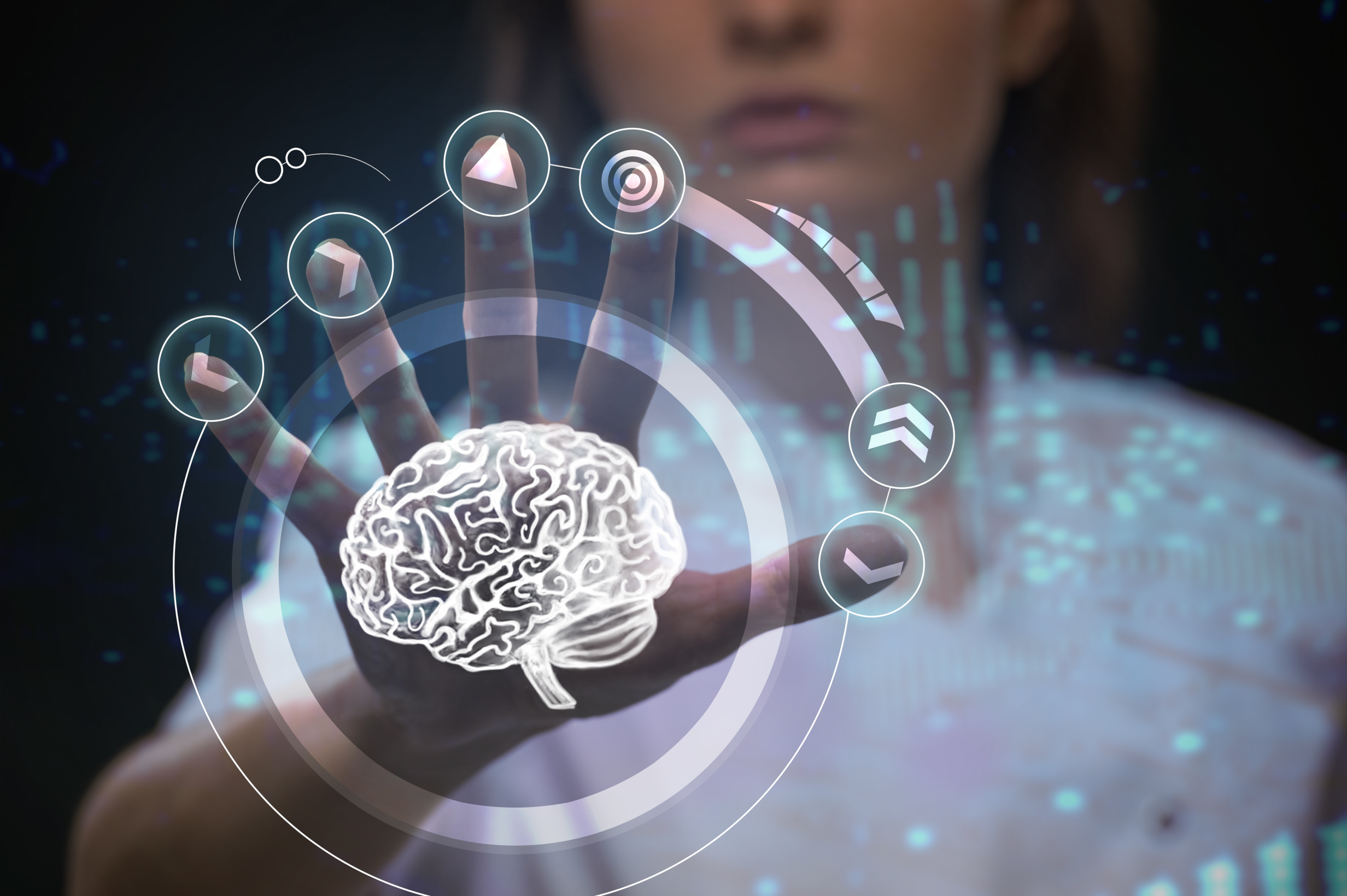
In this case, there is no brain drawn by a hand, but a hand that can handle the brain. This handling is possible thanks to three moves: 1) placing an undetermined background to highlight the brain [Campbell et al., 2015]; 2) separating the brain from the body constructing an organ easy to handle [Martínez Medina, 2021]; and 3) ignoring the brain’s interior by focusing on its external characteristics leaving it as an empty shell even easier to maneuver [Belsky, 2021; Fleck, 1981]. In other words, using simplified drawings makes the brain a clean, transparent, and tame entity [van Dijck, 2005].
4.3 Beyond the flesh: “Matrix brains”
Along with MRI images, the MoCA Cognition website incorporates many other digital brain representations. These images take several forms but often combine digital environments, links, and nodes. In this section, we call these computer-like displays “Matrix brains”, purposefully invoking images from science fiction movies that have the potential to inspire scientific representations [Campbell et al., 2015]. Considering this, we seek to emphasize the remarkable parallels between the Matrix movies [Wachowski & Wachowski, 1999, 2003a, 2003b] and some of the images on the website analyzed in this paper.
The Matrix is a series of films directed by the Wachowski sisters between 1999 and 2003.1 The films take place in a postapocalyptic society in which machines have conquered humans, utilizing them as batteries. Machines connect humans to a virtual reality called the Matrix to keep them mentally active and harvest energy. Hence, the movies occur between the actual reality and this parallel world. Within this framing, the story follows Neo’s odyssey to save Zion, the last human city. Initially, Neo has difficulties distinguishing between the actual reality and the Matrix since both feel real to him. Nevertheless, once he embraces his identity as the “chosen one”, his perception changes. When Neo is in the actual reality, he sees people and things in their material form; when he is in the Matrix, he sees digital entities as chains of green, flowing binary language against a black background. Nevertheless, there is a plot twist at the end. In a battle against Agent Smith, one of the main antagonists, Neo loses his eyes, yet he can see the world in binary language, which suggests that, by losing his fleshy sight, he can see an essence beyond matter.
The Matrix brains on the MoCA Cognition website are used in the main promotional video and the training video. In the main promotional video, it appears when one of the latest MoCA applications is introduced. The background is black, and some blue chains made of 0s and 1s are moving through it. In front of this background is a blue, shiny, translucent brain made of circuits, links, and nodes occupying half of the take. Likewise, these circuits are constantly enlightened as if information flowed through them. Figure 3 is very similar to this, but it misses the binary language.
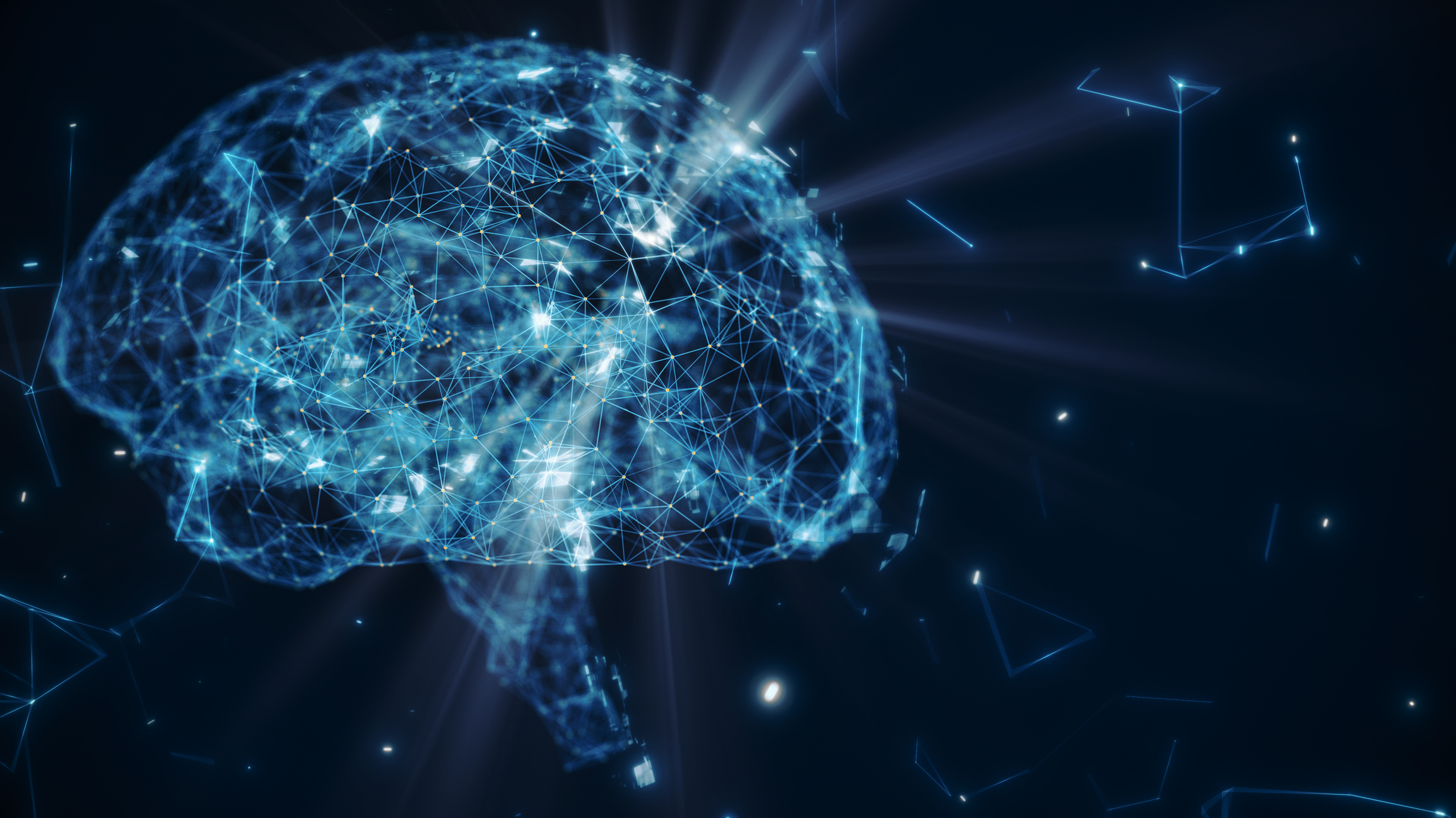
As described with drawings, the image of an isolated floating brain neglects the rest of the body, reflecting the Cartesian divide of body and mind. Similarly, the matrix brain is emptied of its original matter and does not contain structures like the cerebellum, the cortex, or the amygdala. Instead, its content is replaced with complex electric circuits, which is a reappearance of the metaphor of the brain as a hardware-software interface embraced by part of the cognitive sciences [Martin, 2000]. Moreover, this metaphor points toward one of the most preponderant epistemological assumptions embedded in the MoCA Cognition visuality: cognitive domains are comparable to software systems and computers. Then, like the Matrix perception style, once the flesh is disposed of, the essence of the cognitive domains is easier to observe when coded in binary language.
The Matrix brain portrayed in the training video also has a black background, with blue binary text flowing and sparkling around it. This brain is not isolated, but part of a digital human head made of white links and nodes. The nodes and links of the rest of the head are small, while those in the brain are larger and well-defined. Nodes and links also emerge from the head. They adopt the form of capillary structures that go slightly out of the head’s contours as if they could connect with its exterior.
Just as the binary composition of the Matrix movies and the previous Matrix brain, this brain was placed on a black background that renders it more visually striking. Campbell et al. [2015] describe this type of setting as an “extra-planetary space” that erases the presence of other entities to exert control over reality, hiding any political or contextual reflection. In this sense, the Matrix brains on the MoCA Cognition website are situated in a virtual space that overlooks environmental, social, and biological considerations, thereby accentuating their potential for translation via computational means.
Finally, we would like to emphasize the relationship between the Matrix brain and its environment. Brains and bodies are not isolated but semipermeable entities constituted of people, technologies, and things [Mol & Law, 2004; Wolf-Meyer, 2020]. Then, even if the MoCA Cognition reduces and simplifies the space where they are placed, another space is created. Like the Matrix series, the reality around these brains is coded as digital binary language. Furthermore, these brains are not enclosed; they interact with the binary language around them through their capillaries. In this way, they are part of a broader digital environment.
4.4 Cyborg patients: half face-half digital brains
Like the Matrix brains, the MoCA Cognition website uses another fascinating figure: the cyborg patient. According to Donna Haraway [2016], cyborgs are simultaneously machines and animals that blur the boundaries between society and nature, the physical and the nonphysical, mind and body, and symbols and artifacts. Inspired by Haraway, we adopt the term “cyborg patients” to address a particular set of images in which human faces and digital brains are interconnected. In this section, we tackle the cyborg patients in the training video and the news bar of the homepage.
First, the cyborg patient in the training video. The image unfolds with few colors, almost as a black and white take. The background is painted with plain, light gray. In this context, a human face is juxtaposed with a digital brain, as if they were part of the same being. The face is smaller than the brain and presented on a dark gray scale, suggesting to the audience that it does not have a leading role in the setting. Conversely, the brain shines and occupies a more significant part of the frame. It is composed of small white dots and lines that fill its interior and form a net that delimits its contours. Parallelly, colorful prisms made of nodes and links grow from it, covering part of the face.
In the previous section, we discussed the disembodied ontology of the Matrix brains. The hybridization of flesh and digital brain adds another layer of complexity as much as it shows how MoCA Cognition works around the body’s materiality. This brain inhabits a human head, recognizing its connection with a body that sustains it. Yet, this head is secondary in terms of this digital brain’s size, brightness, and liveliness. This situation is accentuated when contemplating the prisms that make the brain’s reach bigger while hiding the face even more, as if the brain is swallowing it.
The second hybrid patient is located on the news bar. It contains three elements: a human face, a digital network, and a finger. The human face is placed on the right side of the frame. At the back of the face is a network composed of bright nodes and links. On the left side, a finger touches the network as if it is entering the interior of the head’s face.
The enactment of bodies in the medical sciences involves body parts, actions, technologies, technical knowledge, and other entities [Belsky, 2021; Daza-Cardona et al., 2021; Fleck, 1981; Martínez Medina, 2021; Mol, 2002; Mol & Law, 2004]. Accordingly, the inclusion of fingers, digital networks, and faces denotes how MoCA Cognition understands knowledge relationships. In this context, the face acts as the patient, the digital network fulfills the role of the brain/cognition encoded as a readable entity, and the finger performs the role of the tester, someone who can touch/know the interior of the patient’s head employing the MoCA. Moreover, the images of cyborg patients discussed in this section suggest that the MoCA can filter out the fleshy components of the body to focus on the cognitive reality.
4.5 Sectioning brains with colors
The last brain images we examine in this paper are what we call “floating three-dimensional brains”. These brains are utilized exclusively in the material related to the training. All of them follow the same pattern: three-dimensional yellow brains suspended in a vacuum while rotating on their axis. They include the brain cortex and folds, the two hemispheres, the cerebellum, and the medulla oblongata. We focus on the three-dimensional brains shown in the description of each of the MoCA’s subscales during the training to be certified as a MoCA user. Every subscale is explained with slides and displays floating digital brains to indicate the brain sections and pathologies associated with the performance in the subscale.
Two of the slides employed to describe the subscale “cube” serve as an illustration. In this subscale, patients need to copy and draw a three-dimensional cube. The first slide is divided into two parts. There is an image of the cube on the right side and a title stating “Anatomy and Pathologies” on the left. Under this title, there is a floating three-dimensional brain with green and blue portions. Below this brain, there are two small charts with the codes of the colors. The blue chart is followed by the assertion “frontal lobe, frontal and subcortical circuits”. The green chart reads, “Right parietal lobe”. This means that the blue areas correspond to the frontal lobe and the green areas to the parietal lobe. The second slide follows a similar scheme. The cube is on the right, and the colored brain is on the left. The only difference is that, instead of codes, there is a list of diseases associated with difficulties completing the task: “Alzheimer’s disease”, “frontal-subcortical vascular disease”, “frontotemporal dementia”, and “Lewy body dementia”.
The setting we just described generates clear delimitations of the brain lobes. This is a resource used in textbooks with neuroanatomical illustrations and digital neuroimaging, in which the brain structures look like discrete entities by using colors and artful work. In this regard, the three-dimensional floating brains encourage a way of looking with the underlying idea that the brain sections are easy to distinguish [Carusi & Hoel, 2015; Casini, 2021; Dumit, 2014]. Nevertheless, the observer who has had the opportunity to see live brains during surgery would note that the lobes are not disconnected and that their color is quite similar.
5 Discussion
In this paper, we conducted a visual analysis of the work done by the brain images on the MoCA Cognition website. Each brain image on this website performs a rhetorical, epistemological, or ontological work. Moreover, rhetoric, epistemology, and ontology are intertwined in each of these images. Let us reflect on this with the first image from the results, which illustrates the relationship between MRIs and the MoCA. By staging an MRI alongside the cognitive domains the MoCA can measure, the image suggests that the MoCA is a valid, updated, and scientific tool (rhetorical work). At the same time, it implies that this cognitive test is as effective as neuroimaging in revealing the brain (epistemological work) and presents the brain as an entity that can be translated into digital information on a screen (ontological work). Taking this into account, we discuss each dimension separately for analytical purposes.
First, the rhetorical work. The primary goals of the MoCA Cognition website are to convince its visitors about the importance of using the MoCA over other cognitive assessments, entice people to purchase the latest software related to this test, and attract new users. In this context, brain images play a pivotal role as rhetorical devices. The display of brains through MRI images reinforces the MoCA’s scientific authority as an objective and reliable test [Carusi & Hoel, 2015]. This is especially powerful when MRI images are subtly incorporated into the website’s background, since, as mentioned, rhetorical strategies are more effective when they go unnoticed [Sismondo, 2009]. Yet, this website does not rely solely on scientificity to capture users’ attention. Scientific websites with marketing aspirations are more flexible in presenting their messages than more formal media [Erikainen et al., 2020]. Therefore, including digital images of colorful brains, cinematic-like takes, and futuristic assemblages contributes to establishing the idea that the MoCA test is a high-tech device and an updated tool.
Second, the epistemological work. The brain images on the studied website revolve around bridging the gap between the cognitive domains and the brain by utilizing the MoCA test. However, brains and cognitive domains are related but not the same. Yet, this website does not visibly explain how they are connected or correlated. Instead, this is implied by showing the brain regions associated with the cognitive domains assessed by the MoCA test. The suggestion is that the brain becomes knowable and accessible through the use of the MoCA exam [Carusi & Hoel, 2015; van Dijck, 2005]. Moreover, some of these images indicate that the cognitive domains assessed by the MoCA test can be translated into numerical values and digital codes. In other words, the MoCA enables one to understand the brain and translate its functions into quantitative measures.
Third, the ontological work. We follow different authors associated with the ontological turn when they propose that every new representation transforms the represented objects [Law, 2004; Martínez Medina, 2016, 2021; Mol, 2002]. Thus, the brain images we analyzed transform the brains themselves. In this sense, we want to highlight that the drawings, the floating three-dimensional brains, and the matrix brains suggest that brains are clean, independent, separable, and controllable [Dumit, 2004, 2014; Fleck, 1981; Martínez Medina, 2021]. Hence, the MoCA Cognition website overlooks the messiness of living brains avoiding their blood irrigation, the color and shape similarities of their components, the skull and the nervous systems that support them, and the physical, affective and social environments in which they are immersed [Wolf-Meyer, 2020]. Furthermore, although the section on cyborg patients illustrates how this website suggests understanding humans as hybridizations between flesh, hardware, and software, it gives precedence to the digital components.
Once the work done by the images on the MoCA Cognition website becomes clear, we want to reflect on how cognition is visualized within a broader cultural context [van Dijck, 2005]. Historically, approaches to understanding cognition have shifted between social psychology and computational sciences. Currently, the excitement around developments in Artificial Intelligence (AI) is driving a resurgence of the latter. This has affected how cognition is depicted on the studied website, as well as in many other health communication media and open-access image databases. These images lead us to solipsistic ideas of the self, personhood, cognition, and intelligence [Dumit, 2004; Rose, 1998].
In this sense, scientific website developers should recognize the effects of the images they post. In our case, MoCA Cognition relies heavily on brain images to tackle cognitive domains. Alternatively, it could post more images of people using their cognitive domains in their everyday lives. This will help visitors understand the cognitive domains in a situated manner, rather than encouraging a disembodied gaze [van Dijck, 2005; Wolf-Meyer, 2020]. These are not superficial decisions. Suppose visitors to neuroscientific websites are encouraged by the images posted there to think that cognitive decline is exclusively located in isolated brains. In that case, they will probably consider individualized and medicalized interventions. In contrast, if they understand the cognitive domains in context, they may consider how people interact with their families, quotidian responsibilities, homes, and neighborhoods; a vision aligned with public health and community interventions.
Building on these insights, we suggest future research to explore how the same brain images can perform different rhetorical, epistemological, and ontological work depending on the observer. This approach considers that images are seen dissimilarly by different audiences at different times and cultural contexts [Awad, 2020; Rose, 2007, 2023; Sturken & Cartwright, 2018]. This research can examine the connections between the communication intended by web designers when placing images on scientific websites and their effects on different visitors. For example, it can be asked: is the work done by images different when researchers, clinicians, policymakers, or patients observe them? How do particular ways of looking affect actual decisions when designing studies, diagnosing patients, constructing health policies, or self-testing one’s own cognitive health? One step in this direction could be studying the users of the most recent MoCA app, Xpresso, which is intended for continuous cognitive self-assessment in people’s households without the direct mediation of health professionals.
Another implication of this paper is its approach to the tensions between rhetoric and science communication. Restrepo Forero [2004] proposes a scientific enterprise that recognizes its own rhetoric and limits by taking responsibility for its persuasive attempts; in other words, a scientific approach that acknowledges its uncertainties, discursive displays, and values. Therefore, websites such as the MoCA cognition can begin by clarifying to their visitors that they are motivated by both scientific practice and profits and that their promotional materials employ simplified representations of the brain. This is particularly relevant given the increasing contention over fake news, post-truth, and trust involved in current scientific communication [Trench, 2025].
Lastly, we urge policymakers and universities to strengthen the education of professional and non-professional audiences when consuming online visual scientific information, especially on private websites. This education should foster a more modest view of science and technology by recognizing the limitations of its promises and questioning the links between high-tech digital representations and progress. For instance, audiences should acknowledge the advantages and disadvantages of cognitive assessment without endorsing the use of cinematic displays.
6 Conclusion
Brain images do all kinds of work in science, education, marketing, and social media. At times, they are playful and humorous; at others, they denote objective truths and are promoted as evidence of the interiority of human minds. They are displayed to symbolize injury, health, intellect, and memory, and are often employed disconnected from the nervous system and body. These disembodied brain images circulate widely, and in this paper, we considered their work on the website of a biomedical tool for measuring cognitive decline and memory impairments.
We understand science communication and the brain images posted on neuroscientific websites as science in the making, with the capacity to shape how brains and cognitive domains are known and seen [Horst et al., 2017; Latour, 1987]. In this vein, we identified that the studied website prioritizes brain images to picture cognition by displaying MRI, drawings, and diverse digital representations. We also explained how these images are used to support the MoCA’s position as a scientific tool and high-tech device in front of current and potential users. These images reinforce the idea that the MoCA test is a preponderant instrument for knowing the brain and the cognitive domains. However, along this path, the brain was portrayed as a simple, transparent entity, obscuring its sociomaterial dimensions.
Finally, we suggest that the images on the studied website and other health communication media should be chosen with more than just aesthetics or credibility in mind. For example, they could incorporate alternative ways to represent cognitive realities in a more contextual manner. Additionally, these media need to be aware of their own rhetorical strategies. On the studied website, the use of digital brain images can be an attractive feature that aligns with the social imagination about brains, AI, and objectivity, but this can also mislead users about the scope of the test it promotes.
Acknowledgments
This research was supported by the Social Sciences and Humanities Research Council of Canada, Grant # 435-2022-0740.
References
-
Abou Elmaaty, A. A., Flifel, M. E., & Zarad, C. A. (2019). Correlation between brain magnetic resonance imaging, cognitive dysfunction and physical dysability in multiple sclerosis. The Egyptian Journal of Neurology, Psychiatry and Neurosurgery, 55(1), 54. https://doi.org/10.1186/s41983-019-0100-0
-
Awad, S. H. (2020). The social life of images. Visual Studies, 35(1), 28–39. https://doi.org/10.1080/1472586x.2020.1726206
-
Beaulieu, A. (2001). Voxels in the brain: neuroscience, informatics and changing notions of objectivity. Social Studies of Science, 31(5), 635–680. https://doi.org/10.1177/030631201031005001
-
Belsky, D. D. (2021). ‘Mutant fish only’: epistemic hybridity and the boundary work of medical illustrators. Sites: a Journal of Social Anthropology and Cultural Studies, 18(2). https://doi.org/10.11157/sites-id492
-
Bloor, D. (1993). Knowledge and social imagery (2nd ed.). The University of Chicago Press.
-
Campbell, N., Deane, C., & Murphy, P. (2015). Advertising nanotechnology: imagining the invisible. Science, Technology, & Human Values, 40(6), 965–997. https://doi.org/10.1177/0162243915574867
-
Carusi, A., & Hoel, A. S. (2015). Brains, windows and coordinate systems. In A. Carusi, A. S. Hoel, T. Webmoor & S. Woolgar (Eds.), Visualization in the age of computerization. Routledge. https://doi.org/10.4324/9780203066973
-
Casini, S. (2021). Giving bodies back to data: image makers, bricolage, and reinvention in magnetic resonance technology. The MIT Press.
-
Casini, S., Cati, A., & Toschi, D. (2022). Sick and injured bodies: medical imagery and media practices of care. Cinéma & Cie. Film and Media Studies Journal, 22(39), 9–20. https://doi.org/10.54103/2036-461x/19083
-
Daza-Cardona, J. A., Vargas-Ramírez, J., & Guapacha-Sánchez, M. A. (2021). Doing odontograms and dentists in the classroom. Materiality and affect in dental education. Tapuya: Latin American Science, Technology and Society, 4(1), 1968635. https://doi.org/10.1080/25729861.2021.1968635
-
de Boer, B., te Molder, H., & Verbeek, P.-P. (2020). Constituting ‘visual attention’: on the mediating role of brain stimulation and brain imaging technologies in neuroscientific practice. Science as Culture, 29(4), 503–523. https://doi.org/10.1080/09505431.2019.1710739
-
Dumit, J. (2004). Picturing personhood: brain scans and biomedical identity. Princeton University Press. https://doi.org/10.1515/9780691236629
-
Dumit, J. (2014). How (not) to do things with brain images. In C. Coopman, J. Vertesi, M. Lynch & S. Woolgar (Eds.), Representation in scientific practice revisited (pp. 291–314). The MIT Press. https://doi.org/10.7551/mitpress/9780262525381.003.0014
-
Erikainen, S., Couturier, A., & Chan, S. (2020). Marketing experimental stem cell therapies in the UK: biomedical lifestyle products and the promise of regenerative medicine in the digital era. Science as Culture, 29(2), 219–244. https://doi.org/10.1080/09505431.2019.1656183
-
Fähnrich, B., Weitkamp, E., & Kupper, J. F. (2023). Exploring ‘quality’ in science communication online: expert thoughts on how to assess and promote science communication quality in digital media contexts. Public Understanding of Science, 32(5), 605–621. https://doi.org/10.1177/09636625221148054
-
Fleck, L. (1981). Genesis and development of a scientific fact (F. Bradley & T. J. Trenn, Trans.). The University of Chicago Press.
-
Geller, R., & Slicer, K. (2024). Montreal Cognitive Assessment (MoCA). In C. J. Golden & R. Bennett (Eds.), Clinical integration of neuropsychological test results (pp. 191–204). CRC Press. https://doi.org/10.1201/9781003309604
-
Gruber, D. (2021). Neurons in sparkling space: scientific objectivity and ‘blurry’ images in neuroscience. JCOM, 20(01), A02. https://doi.org/10.22323/2.20010202
-
Haraway, D. (2013). The persistence of vision. In N. Mirzoeff (Ed.), The visual culture reader (3rd ed., pp. 356–362). Routledge.
-
Haraway, D. (2016). A cyborg manifesto: science, technology, and socialist-feminism in the late twentieth century. In Manifestly Haraway (pp. 3–90). University of Minnesota Press.
-
Horst, M., Davies, S. R., & Irwin, A. (2017). Reframing science communication. In U. Felt, R. Fouché, C. A. Miller & L. Smith-Doerr (Eds.), The handbook of science and technology studies (4th ed., pp. 881–907). The MIT Press.
-
Joyce, K. A. (2006). From numbers to pictures: the development of magnetic resonance imaging and the visual turn in medicine. Science as Culture, 15(1), 1–22. https://doi.org/10.1080/09505430600639322
-
Khalili-Mahani, N., & Loos, E. (2023). A critical perspective on the mediatization of brain imaging and healthy ageing. JCOM, 22(05), Y01. https://doi.org/10.22323/2.22050401
-
Latour, B. (1987). Science in action: how to follow scientists and engineers through society. Harvard University Press.
-
Law, J. (2004). After method: mess in social science research. Psychology Press. https://doi.org/10.4324/9780203481141
-
Mantilla, M. J. (2018). La difusión de las neurociencias en Argentina: un análisis de las motivaciones de los neurocientíficos para la comunicación pública de la ciencia. Redes: Revista de Estudios Sociales de la Ciencia y la Tecnología, 24(46), 87–104. https://doi.org/10.48160/18517072re46.90
-
Martin, E. (2000). Mind-body problems. American Ethnologist, 27(3), 569–590. https://doi.org/10.1525/ae.2000.27.3.569
-
Martínez Medina, S. (2016). Hacer arteria carótida en el Laboratorio de Anatomía. Práctica y materialidad en una asignatura de Medicina. Revista Colombiana de Sociología, 39(2), 31–47. https://doi.org/10.15446/rcs.v39n2.58964
-
Martínez Medina, S. (2021). Anatomización. Una disección etnográfica de los cuerpos. Universidad de los Andes. https://doi.org/10.30778/2021.02
-
Martínez-Medina, S. (2022). Knowledge practices. In H. Callan (Ed.), The International Encyclopedia of Anthropology. John Wiley & Sons. https://doi.org/10.1002/9781118924396.wbiea2516
-
McCabe, D. P., & Castel, A. D. (2008). Seeing is believing: the effect of brain images on judgments of scientific reasoning. Cognition, 107(1), 343–352. https://doi.org/10.1016/j.cognition.2007.07.017
-
Mol, A. (2002). The body multiple: ontology in medical practice. Duke University Press. https://doi.org/10.1215/9780822384151
-
Mol, A., & Law, J. (2004). Embodied action, enacted bodies: the example of hypoglycaemia. Body & Society, 10(2–3), 43–62. https://doi.org/10.1177/1357034x04042932
-
Ortega, L., & Nirappil, F. (2024, July 9). What cognitive tests measure and could tell us about Biden and Trump. The Washington Post. https://www.washingtonpost.com/health/2024/07/09/cognitive-test-moca-biden-trump/
-
Petchesky, R. P. (1987). Fetal images: the power of visual culture in the politics of reproduction. Feminist Studies, 13(2), 263–292. https://doi.org/10.2307/3177802
-
Pinch, T. J. (1993). La retórica y la controversia sobre la fusión fría: del Woodstock químico al altamont físico. Política y Sociedad, 14/15, 155–170. https://revistas.ucm.es/index.php/POSO/article/view/POSO9394110155A
-
Restrepo Forero, O. (2004). Retórica de la ciencia sin retórica. Revista Colombiana de Sociología, 23, 251–268. https://revistas.unal.edu.co/index.php/recs/article/view/11280
-
Rollins, N., Piwoz, E., Baker, P., Kingston, G., Mabaso, K. M., McCoy, D., Ribeiro Neves, P. A., Pérez-Escamilla, R., Richter, L., Russ, K., Sen, G., Tomori, C., Victora, C. G., Zambrano, P., & Hastings, G. (2023). Marketing of commercial milk formula: a system to capture parents, communities, science, and policy. The Lancet, 401(10375), 486–502. https://doi.org/10.1016/s0140-6736(22)01931-6
-
Rose, G. (2007). Visual methodologies: an introduction to the interpretation of visual materials (2nd ed.). SAGE Publications.
-
Rose, G. (2023). Visual methodologies: an introduction to researching with visual materials (5th ed.). SAGE Publications.
-
Rose, N. (1998). Inventing our selves: psychology, power, and personhood. Cambridge University Press. https://doi.org/10.1017/CBO9780511752179
-
Sismondo, S. (2009). Ghosts in the machine: publication planning in the medical sciences. Social Studies of Science, 39(2), 171–198. https://doi.org/10.1177/0306312708101047
-
Sturken, M., & Cartwright, L. (2009). Practices of looking: an introduction to visual culture (2nd ed.). Oxford University Press.
-
Sturken, M., & Cartwright, L. (2018). Practices of looking: an introduction to visual culture (3rd ed.). Oxford University Press.
-
Trench, B. (2025). Trust and mistrust in science: beyond the binary. In A. Fage-Butler, L. Ledderer & K. H. Nielsen (Eds.), Science communication and trust (pp. 323–343). Springer. https://doi.org/10.1007/978-981-96-1289-5_16
-
van Dijck, J. (2005). The transparent body: a cultural analysis of medical imaging. University of Washington Press.
-
Wachowski, L. and Wachowski, L. (Directors). (1999). The Matrix [Film]. Warner Bros.
-
Wachowski, L. and Wachowski, L. (Directors). (2003a). The Matrix Reloaded [Film]. Warner Bros.
-
Wachowski, L. and Wachowski, L. (Directors). (2003b). The Matrix Revolutions [Film]. Warner Bros.
-
Wolf-Meyer, M. (2020). Neurological disorders, affective bioethics, and the nervous system: reconsidering the Schiavo case from a materialist perspective. Medical Humanities, 46(3), 166–175. https://doi.org/10.1136/medhum-2018-011568
-
Zhang, S., Kwapong, W. R., Yang, T., Liu, P., Tuo, Q., Cheng, Y., Li, X., Liu, M., Lei, P., & Wu, B. (2021). Choriocapillaris changes are correlated with disease duration and MoCA score in early-onset dementia. Frontiers in Aging Neuroscience, 13, 656750. https://doi.org/10.3389/fnagi.2021.656750
Notes
1. We do not consider the 2021 Matrix release for our analysis because the plot takes a different direction from the original trilogy.
About the authors
Jorge Daza’s research is at the intersection of Science and Technology Studies and Health Sciences. He has been an investigator in projects related to visual culture in neurosciences, scientific training in psychiatry and dentistry, health participation, and forced displacement. He has taught in medicine, dentistry, psychiatry, and psychology programs in courses about social research, community projects, epistemology, and clinical interventions in Colombia. He has a BS in Psychology, an MSC in Social Studies of Science, and an MA in Science and Technology Studies. Currently, he is doctoral student in Science and Technology Studies at York University. He has published articles in Trilogía Ciencia Tecnología Sociedad, Tapuya, and other journals.
E-mail: daza674@gmail.com
Denielle Elliott, Ph.D., is a Professor of socio-cultural anthropology at York University, where she holds a York Research Chair in Injured Minds. She is also the Deputy-Director of the Institute for Technoscience and Society. Her current research explores the embodied experience of people living with altered neuro states and knowledge-making practices in the clinic and laboratory, and their convergences. She is particularly interested in how patients, doctors, and scientists make sense of that which is imperceptible and often beyond language and scientific knowing. As part of this, her research considers how sensory ethnographic methods can augment neuro-scientific understanding and facilitate collaborative and empowering epistemological approaches in neurology.
E-mail: dae@yorku.ca